By A Mystery Man Writer

Eguides Shoulder – Crossover Symmetry

Hassan KESSERWANI, Consultant, Doctor of Medicine, Neurology

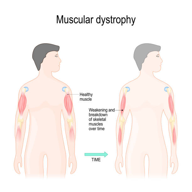
Whole-muscle fat analysis identifies distal muscle end as disease initiation site in facioscapulohumeral muscular dystrophy

MRI findings of chronic distal tendon biceps reconstruction and associated post-operative findings
Atrophied arm hi-res stock photography and images - Alamy

Identification of novel FHL1 mutations associated with X-linked scapuloperoneal myopathy in unrelated Chinese patients

Post-Polio-Like Syndrome The American Journal of Medicine Blog

Hassan KESSERWANI, Consultant, Doctor of Medicine, Neurology
Muscle atrophy hi-res stock photography and images - Alamy
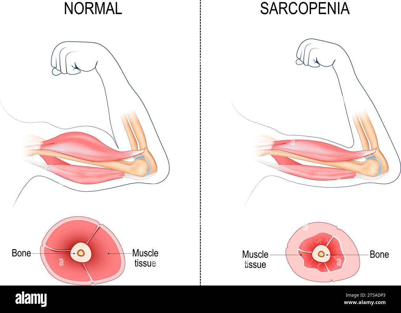
90+ Muscle Atrophy Stock Illustrations, Royalty-Free Vector Graphics & Clip Art - iStock

Axillary nerve entrapment by paralabral cyst
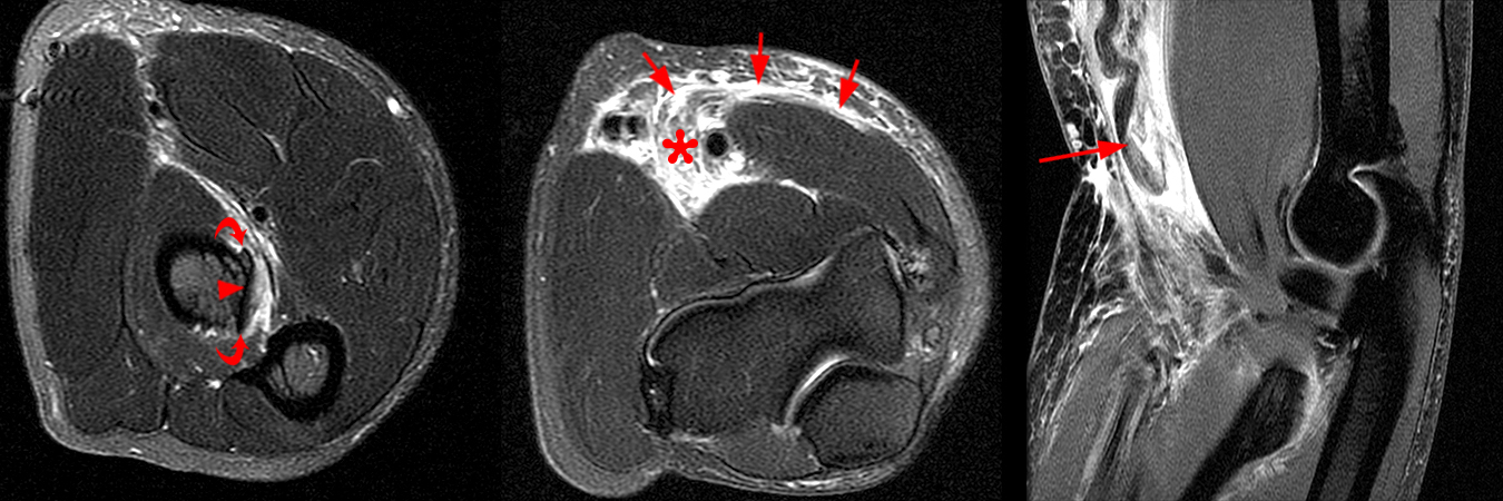
Distal Biceps Tendon Rupture Elbow
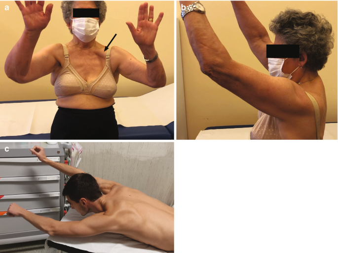
Neuropathies and Nerve Entrapments Around the Scapula and the Shoulder

Physiological and pathological skeletal muscle T1 changes quantified using a fast inversion-recovery radial NMR imaging sequence