
Download scientific diagram | (A) A brightness mode (b-mode) image of the lateral abdominal wall. Abbreviations: EO, external oblique; IO, internal oblique; TrA, transversus abdominis. (B) A split-screen image with b-mode on the left and motion mode (m-mode) on the right. The m-mode image represents the information from the dotted line on the b-mode image displayed over time (x-axis). Static structures produce straight interfaces while structures that change in thickness or depth (in this case the TrA) create curved interfaces. The increase in depth of the TrA correlates to a contraction. Reproduced with permission Whittaker 2007. 142 from publication: Rehabilitative Ultrasound Imaging: Understanding the Technology and Its Applications | The use of ultrasound imaging by physical therapists is growing in popularity. This commentary has 2 aims. The first is to introduce the concept of rehabilitative ultrasound imaging (RUSI), provide a definition of the scope of this emerging tool in regard to the physical | Rehabilitation, Ultrasonography and Ultrasound Imaging | ResearchGate, the professional network for scientists.
Basic ultrasound

Providing Visual Biofeedback Using Brightness Mode Ultrasound During a Golf Swing

Brightness mode ultrasound (B-mode): grayscale ultrasound showing fiber
Fundamentals of transesophageal echocardiography (Section 1) - Core Topics in Transesophageal Echocardiography
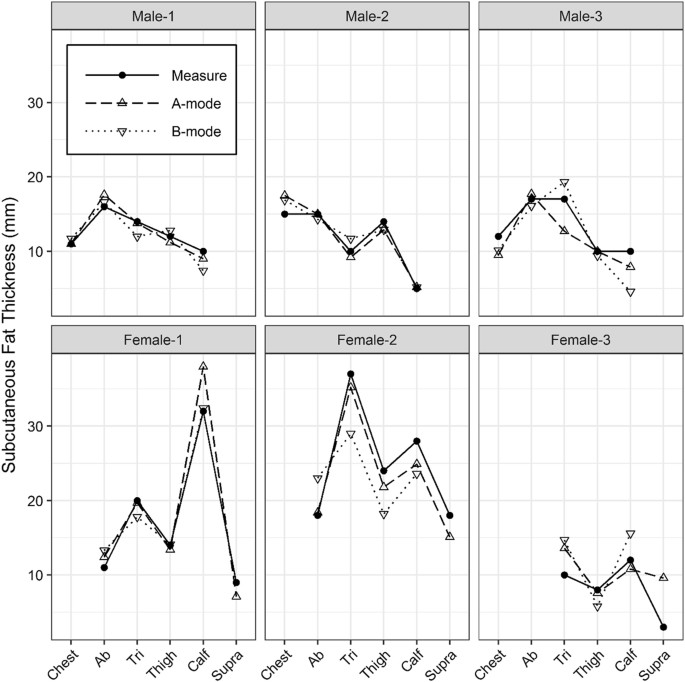
A-mode and B-mode ultrasound measurement of fat thickness: a cadaver validation study

Measurement of linea alba distortion and linea alba stiffness. (A)

IJERPH, Free Full-Text
Point-of-Care Ultrasound for Outpatient Neurology - Practical Neurology

A-mode and B-mode ultrasound measurement of fat thickness: a cadaver validation study
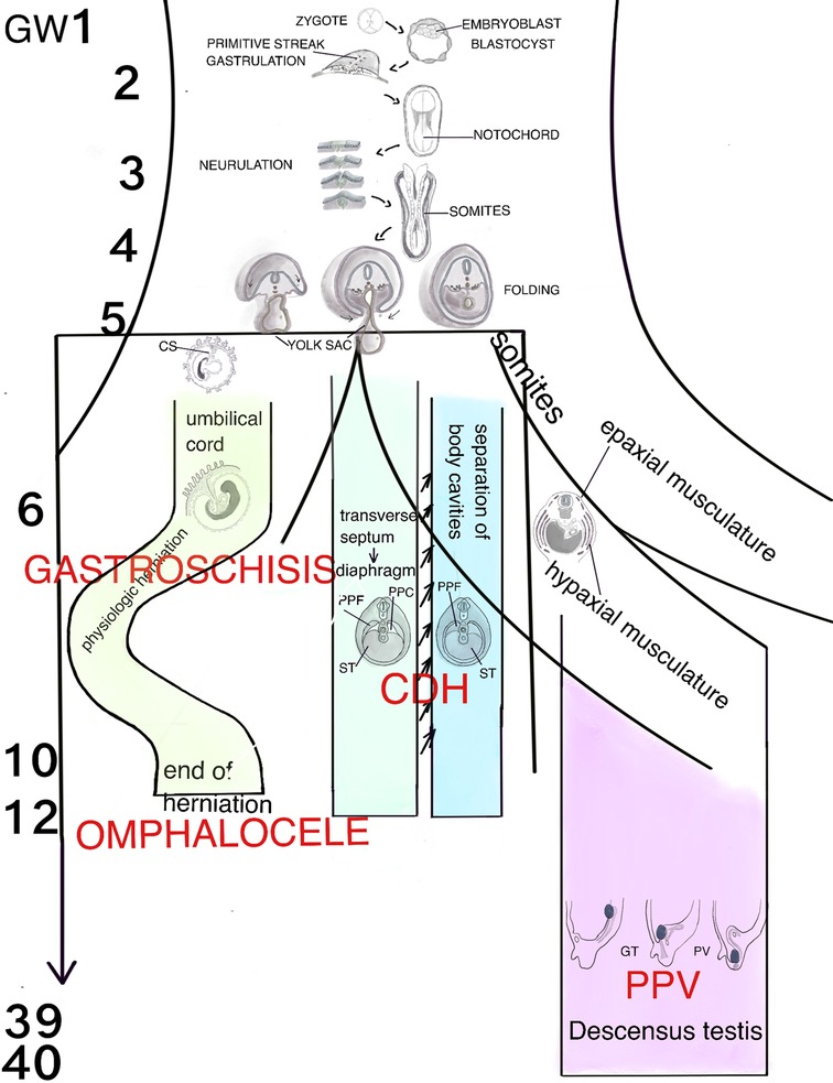
Frontiers Embryology of the Abdominal Wall and Associated Malformations—A Review

PDF) Rehabilitative Ultrasound Imaging: Understanding the

A) A brightness mode (b-mode) image of the lateral abdominal wall.

Medical ultrasound - Wikipedia
Basics of Ultrasound: Pitfalls and Limitations - NYSORA